
Transcript
Announcer:
You’re listening to Eye on Ocular Health on ReachMD, and this episode is sponsored by Apellis Pharmaceuticals, Inc. Here’s your host, Dr. Alexandria May.
Dr. May:
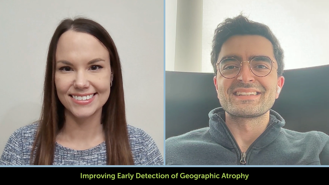
Welcome to Eye on Ocular Health on ReachMD. I'm Dr. Alexandria May, and joining me to discuss the early detection and management of geographic atrophy is Dr. Hasenin Al-khersan. He's a board-certified medical and surgical retina specialist with Retina Consultants of Texas in Houston. Dr. Al-khersan, thanks so much for being here today.
Dr. Al-khersan:
Thanks so much for having me. I'm really looking forward to our conversation.
Dr. May:
To start us off, Dr. Al-khersan, where do you see the biggest challenges when it comes to catching geographic atrophy early, before significant vision loss occurs?
Dr. Al-khersan:
The biggest challenge with geographic atrophy is that it can progress, and the patient might not know about it. So the way that geographic atrophy works is, it usually has the biggest impact on vision the closer it moves to the fovea in the center of the macula. But if it starts to grow in the peripheral parts of the macula, it may be doing damage without the patient initially knowing.
I like to use the analogy of glaucoma, which is very similar. In the early stages, the patient may not know that they have glaucoma, and by the time they start having visual symptoms, it can be too late, in the sense that the type of damage that's done to the retina is irreversible—at least with the current therapies that we have now.
It's required a little bit of retraining, not only for referring physicians, but also for retina specialists, because historically, we didn't have any treatments for geographic atrophy. So you wanted to identify it, but if you didn't, it wasn't as big of a deal, because we just didn't have any treatment for it at all.
Now, that's changed. We have two approved complement-based therapies, and they both tend to work better if they're initiated earlier, based on the trial data that we have available. And so it's gone from not being that big of a deal if you don't identify geographic atrophy in terms of the consequences to having more consequences now for the patients’ potential to preserve their retina.
And so I think that paradigm shift is one of the biggest challenges: the fact that it progresses unbeknownst to the patient in many cases, and the fact that it's irreversible, as well.
And then the other important thing that I would say is that it can be difficult to risk stratify patients. Not every patient with intermediate AMD, or even early geographic atrophy, is going to behave the same. Some patients are going to progress more quickly than others. And until recently, we didn't really have a great sense of how to stratify patients into different cohorts based on how they might progress, and so that's another challenge as well.
Dr. May:
Let's talk about optical coherence tomography, or OCT. What are the specific structural findings that are shifting how we think about early detection? And what are clinicians potentially missing if they're relying solely on conventional imaging?
Dr. Al-khersan:
Historically, we just had color fundus photography, and we would try to grade geographic atrophy or AMD solely based on that. Then, we progressed to fundus autofluorescence, which was a little bit better, because it was a little bit clearer, and we could see where there were areas of death of the photoreceptors in the retinal pigment epithelium, or the RPE.
But OCT is, by far, the most rigorous tool that we have available to us. It allows us to look at the tissue layers in really high resolution—a resolution that can't be achieved with fundus autofluorescence or color fundus photographs alone. And so, going back to what we were saying before, it's much easier to pick up changes before they really start having big effects on the patient's vision.
So some of the most important features of intermediate AMD and subsequently, geographic atrophy, are the loss of the retinal pigment epithelium. That's kind of the last stage. That's really when the retina is basically starting to die off. But there are some features that you can see before that. Certainly, the amount and type of drusen—little deposits underneath the retina—can signal a likelihood to progress. We've identified something called subretinal drusenoid deposits as a high-risk feature. So these types of drusen sit underneath the retina and between the RPE, and they can be a signal that a patient might be more likely to progress. And they're best characterized on OCT. So that's one of the important features. There's something called hyperreflective foci. They're the little bright spots within the retina, that, again can be identified on the OCT. They're very distinctive and easy to see there.
And then, as I mentioned, I tell my patients OCT is like looking at a cake. We can see each distinct layer of the retina. And so not only do we see the RPE, which is what dies off in geographic atrophy, but we can also see damage to photoreceptors or the ellipsoid zone before that happens, and that can give us a sense of how stressed the retina is. And more and more research is coming out now that this ellipsoid zone is very important, and that it can tell us how likely a patient is to progress, particularly if we weigh the amount of damage that’s been done to the ellipsoid zone relative to the amount of damage that's been done to the RPE or the retinal pigment epithelial layer there.
So all of these features are best seen on OCT. Some of them you can sometimes see on color fundus photographs or fundus autofluorescence, but certainly, without OCT, it can be very difficult to pick these findings up.
Dr. May:
And building on those findings, what are some of the key imaging biomarkers that help clinicians identify patients who are at higher risk of progression and foveal threat?
Dr. Al-khersan:
The first of these is very simple: location. The closer a lesion is to the fovea, the more likely that it's going to impair the patient's vision. And then the size of the lesion, of course. Larger lesions mean more tissue is affected and, again, it’s more likely that the patient's vision is affected.
There’s something called circularity. So the less circular a lesion is and the more surface area it has, the faster it's likely to progress. And that just has to do with the fact that these lesions are growing at the borders. So if you have more border, you have more growth. Multifocal lesions tend to grow faster than just single lesions. And that has to do with surface area, as well. You could have the same area spread across different lesions compared to one lesion, and that multifocal lesion will grow faster on autofluorescence. If the lesion has a hyperautofluorescence border, that usually tells you that it's active, as opposed to, if there isn't an increased autofluorescence signal, that may be less likely to progress.
And then all of the features we discussed from the OCT earlier, which include the subretinal drusenoid deposits, the hyperreflective foci, and easy loss as well. And the last thing I'll say—I don't know if it would technically be classified as a biomarker—but the patient's fellow eye status. So if the patient has geographic atrophy that may be more progressed in the other eye, usually one eye will follow the other in terms of the nature of how the GA lesion will progress. And we've seen that in clinical trials, as well. So that can give you a good idea, if the other eye's already been affected, how the eye under study may progress.
Dr. May:
For those just tuning in, you're listening to Eye on Ocular Health on ReachMD. I'm Dr. Alexandria May, and I'm speaking with Dr. Hasenin Al-khersan about identifying geographic atrophy earlier to improve patient outcomes.
So, Dr. Al-khersan, taking this a step further now, how are these imaging findings influencing real-world clinical decisions regarding monitoring intervals and referral timing?
Dr. Al-khersan:
I think it's giving us a more sophisticated understanding in terms of stratifying patients. So if we believe a patient is more likely to progress—let's say they just have intermediate AMD, but they have some of these high-risk features—I might follow them more closely than I would somebody that has none of these features. I think, historically, we've had a pretty protocolized follow-up regimen. But as we get this more sophisticated understanding, we can group patients into different buckets for their follow-up schedules, and then we can intervene earlier and potentially save them from losing vision significantly. And so I think it's that risk stratification that bleeds into our follow-up that is the most important thing.
And then, again, now, in the era where we actually have therapeutics, that becomes much more important than it was ever in the past. Unfortunately, in the past, if you did identify a patient progressing, you really couldn't do anything about it. And that's just not the case anymore.
Dr. May:
With that being said, what should an optimized referral pathway look like for patients who are suspected of progressing towards geographic atrophy? And what role should the referring clinician play in monitoring after the patient is referred?
Dr. Al-khersan:
The way I think about it now, given that there is a therapeutic available, any patient with geographic atrophy should be seeing a retina specialist. And it doesn't necessarily mean that they're going to be treated, but they should be evaluated in the context of how their geographic atrophy looks and whether they have any of these high-risk features. And that decision can be made jointly with the patient.
Earlier on, when the patient has intermediate AMD, I would say any of those findings should trigger a referral, really. Those findings we talked about on the OCT and on the autofluorescence may suggest that this patient may convert to geographic atrophy in the near future.
I tell my referring docs that I'd rather be safe than sorry. And so, if they have concern for a patient, just refer them and have them looked over. And then what I tend to do in terms of the shared care model is if I think a patient is less likely to progress or they don't necessarily need treatment, I'll have them refer back to optometry or the comprehensive eye clinic, where they can continue to be monitored. And then we can alternate visits sometimes. Or if they're really low risk, I just have them follow up with their primary eye care specialist.
Dr. May:
Finally, Dr. Al-khersan, as imaging technologies continue to evolve, where do you see the biggest opportunities to further improve early detection and outcomes?
Dr. Al-khersan:
So this is a labor of love for me. One of my research focuses is on artificial intelligence and the use of automated tools powered by AI to help us detect these features. So it's great to talk about subretinal drusenoid deposits and hyperreflective foci and all this stuff, but it's actually quite difficult to quantify them, to sit in clinic there and scroll through every single OCT B-scan. The reality is, nobody's really doing that. We currently just use the gestalt; if we see it, we flag it in our brain, but we're not really quantifying it. And that's where artificial intelligence is going to make a huge difference.
There are certain regulatory hurdles. We have to get these algorithms approved. We have to make sure they're effective and safe for patients. But that quantification and that automated trigger will allow us to interpret the OCTs in a much quicker fashion than we can do on our own. Especially when we're having a busy clinic, it's hard.
And the other component of that is, one step is identifying them, the second step is seeing any change, because it's the change that's the most important, especially early on in disease. And these programs will allow us to understand when these changes are happening, again, in a much more objective fashion than what we can currently do.
And then the third prong of that is it will allow us to create risk tools, basically, to tell us that question of who is going to progress. Right now we have these biomarkers individually that tell us this patient is more likely to progress than a patient without them. But in the context of all these biomarkers together—potentially the patient's age, obviously their smoking status, whether they use AREDS—can give us a global risk score for an individual patient. It'll deliver a much more personalized medicine approach compared to what we currently do.
Other areas where I think AI will help is in connecting clinical care networks. We have great relationships with our referring docs, but sometimes things get lost in translation. You know, you tell a patient, "Hey, you got to see a retina doc," and they don't go in, or the retina clinic doesn't schedule the appointment. There's a lot of areas where the patient can get lost. But I think, in the future, we'll have, again, automated ways where, if the algorithm picks up that the patient has these findings, it'll let the doc know in the primary eye care clinic, "Hey, this patient should likely be referred." And then it'll go the next step of saying, "You have retina clinic A in this area. Would you like to schedule an appointment for them?" And it can potentially just automatically trigger all of that.
And then one more thing I'd like to point out is home monitoring. So for some patients, it's really a lot to come into clinic, particularly for, these older folks. Obviously, AMD is age-related. And so coming to the clinic can be a huge burden for them and their caregivers. I think we already do have some devices, more on the wet AMD side, that will help us monitor for fluid. But in the future, I think we'll have these tools available to us—AI-powered tools that the patient can just kind of pop their eye in, and then it'll tell them, "Hey, you have some change.
Get into the clinic,” or “You should see your optometrist, ophthalmologist, or retina specialist."
Dr. May:
With those forward-looking insights in mind, I want to thank my guest, Dr. Hasenin Al-khersan, for sharing his expertise on how early detection can shape care for patients with geographic atrophy. Dr. Al-khersan, it was great having you on the program.
Dr. Al-khersan:
Thanks so much for your time. It’s a really important conversation for our patients.
Announcer:
This episode of Eye on Ocular Health was sponsored by Apellis Pharmaceuticals, Inc. To access this and other episodes in our series, visit Eye on Ocular Health on ReachMD.com, where you can Be Part of the Knowledge. Thanks for listening!